SKU: SKU: EZF220
Erler-Zimmer Eye, 6 times size, 6 parts
Verpackungseinheit :
Couldn't load pickup availability


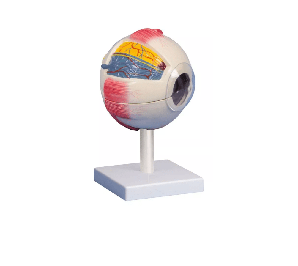
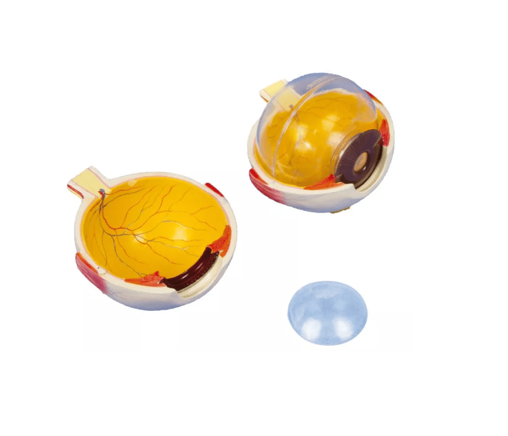
This large-format eye model, magnified 6x, offers an impressively detailed view of the anatomy of the human eye. The model can be opened horizontally to reveal the internal structures of the eyeball —ideal for anatomy teaching, medical training , or patient counseling in ophthalmology.
The cornea, iris, lens, and vitreous body are individually removable, allowing for a detailed understanding of the structure and function of the visual system. Additionally, muscle attachments to the sclera and part of the choroid are depicted. The model is securely mounted on a stand and is ideal for presentations or classroom use.
Areas of application:
-
Training in ophthalmology & optometry
-
Demonstration in class & everyday clinical practice
-
Patient education about eye diseases and operations
Advantages at a glance:
-
6x magnification for maximum clarity
-
Can be disassembled into cornea, iris, lens and vitreous body
-
With visible muscle attachments & choroid portion
-
Stable mounted on a tripod
Further information:
-
Horizontally divisible for optimal insight into the interior of the eye
-
Ideal for illustrating lens function, visual processes & diseases
-
Particularly suitable for visually supported training & consulting
Delivery information:
PZN: " "
WEEE number:
Assistive device number:
Manufacturer:
Erler-Zimmer GmbH & Co. KG
Hauptstrasse 27
77886 Lauf
Deutschland
Tel: +49 07841 / 67191-0
Email: info@erler-zimmer.de
VAT ID number: Achtung! Medizinisches Ausbildungsmaterial, kein Spielzeug. Nicht geeignet für Personen unter 14 Jahren. Attention! Medical training material, not a toy. Not suitable for persons under 14 years of age.