SKU: SKU: EZF50
Erler-Zimmer Half of the eye enlarged
Verpackungseinheit :
Couldn't load pickup availability


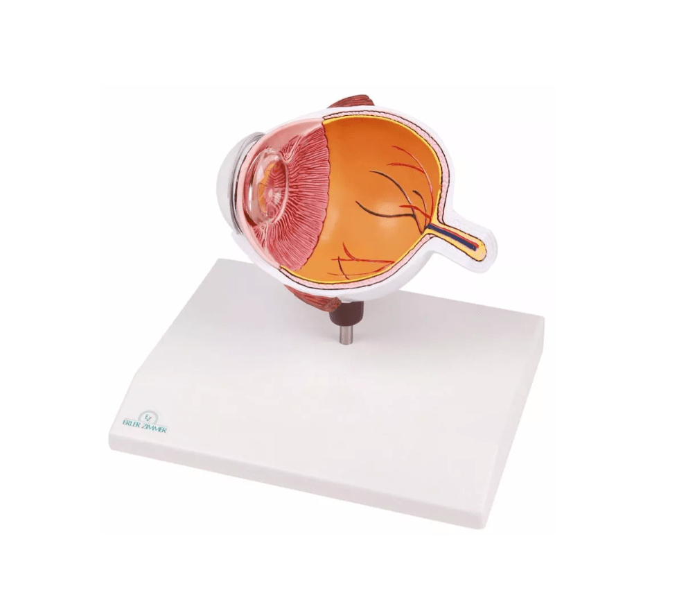
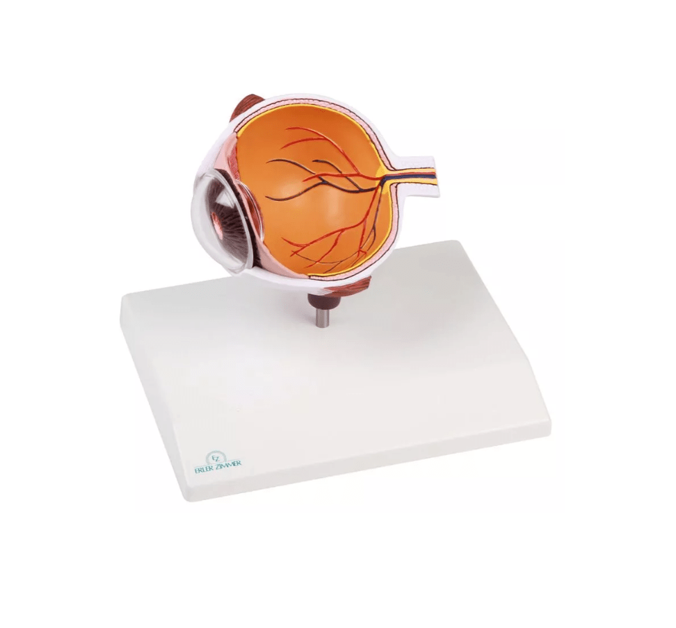
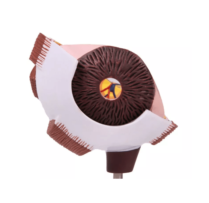
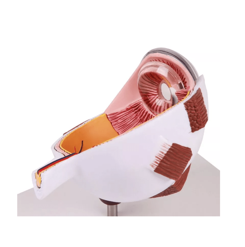
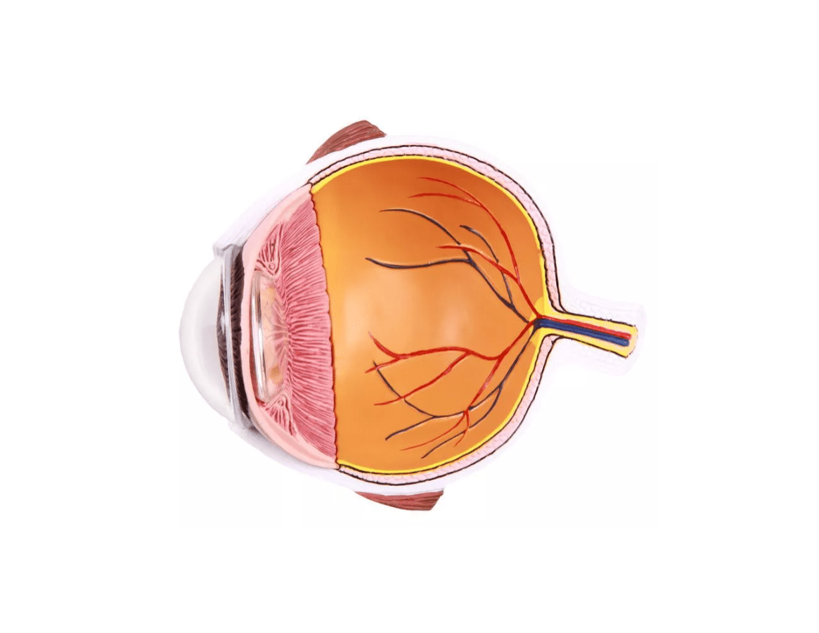
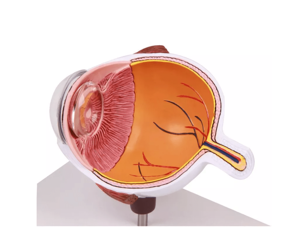
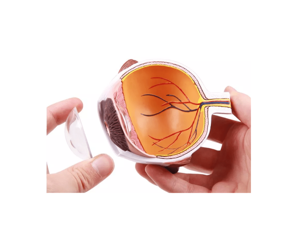
This enlarged cross-sectional model of one half of the eye offers a vivid insight into the most important internal and external structures of the human eye – ideal for anatomy lessons, ophthalmological training or patient counseling .
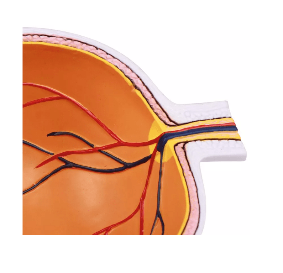
Depicted are, among other things, the choroid, retina, macula, optic nerve head , central retinal artery and vein , as well as the optic nerve and its supplying vessels. The external eye muscles (superior and inferior rectus muscles), the ciliary body , the iris , the sclera , and the cornea are also anatomically correct.
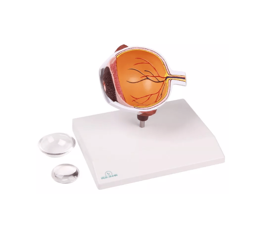
The lens and cornea are removable , allowing additional insights into the internal structure of the eye. The model is part of the EZ Augmented Anatomy series and can be used with the free AR app : This app automatically recognizes the model and displays the nomenclature in augmented reality – anytime, anywhere, and without registration.
Areas of application:
-
Anatomy teaching and training in ophthalmology & nursing
-
Patient education in ophthalmology practice or clinic
-
Complement to digital learning formats with AR function
Advantages at a glance:
-
Illustrative cross-sectional model of one half of the eye
-
Removable lens & cornea to illustrate internal structures
-
Compatible with the EZ Augmented Anatomy app
Further information:
-
Representation of the macula & optic nerve structure
-
Including retinal blood vessels & central artery/vein
-
Supports modern learning through app integration
Delivery information:
PZN: " "
WEEE number:
Assistive device number:
Manufacturer:
Erler-Zimmer GmbH & Co. KG
Hauptstrasse 27
77886 Lauf
Deutschland
Tel: +49 07841 / 67191-0
Email: info@erler-zimmer.de
VAT ID number: Achtung! Medizinisches Ausbildungsmaterial, kein Spielzeug. Nicht geeignet für Personen unter 14 Jahren. Attention! Medical training material, not a toy. Not suitable for persons under 14 years of age.