SKU: SKU: EZMP1121
Erler-Zimmer Pericardial space model
Verpackungseinheit :
Couldn't load pickup availability


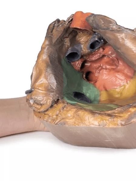

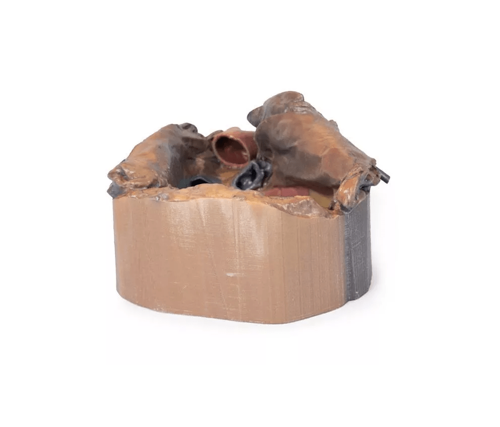

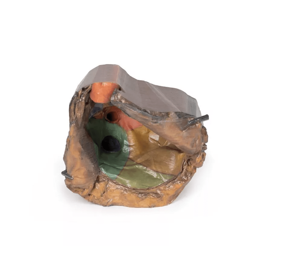
The Pericardial Space Model provides a detailed and precise representation of the anatomical structures of the heart and its pericardium. In this unique specimen, the heart has been removed to demonstrate the reflections of the parietal peritoneum and the orientation of the heart relative to neighboring structures such as the diaphragm and lungs. This innovative representation allows students and medical professionals to grasp the precise relationships between the various components of the heart and pericardium in a clear, understandable manner.
Features and functions:
-
Anatomical detail: The model depicts the precise anatomy of the pericardial space, including the structure of the parietal and visceral pericardium surrounding the heart. The transition from the parietal pericardium to the epicardium (visceral pericardium) is depicted in detail to better understand the interaction of these layers.
-
Heart and its structures: This model demonstrates the base of the heart, which extends superiorly and posteriorly in front of the pulmonary hilum. It shows the atria (left and right) located at this location, as well as the proximal parts of the great vessels such as the aorta and the pulmonary trunk.
-
Clinically relevant structures: The transverse sinus and oblique pericardial sinus are clearly depicted to illustrate their role in specific cardiac surgeries and clinical scenarios. The transverse sinus is visible between the aorta and the pulmonary trunk, while the oblique pericardial sinus is depicted between the pulmonary veins.
-
Great Vessels and Pulmonary Vessels: The model includes detailed representations of the aorta, superior and inferior vena cava, and the pulmonary trunk, including the left and right pulmonary arteries and veins. These structures are clearly visible in their relationship to each other and to the cardiac regions.
-
Diaphragm and lung relationships: The model shows how the heart rests on the diaphragm surface and how the lung surfaces of the heart mirror the left and right lungs. The model also depicts the apex of the heart, located in the left fifth intercostal space.
Advantages:
-
Enhanced learning opportunities: This model provides in-depth visualization of the cardiac pericardial space and surrounding structures. It helps medical students and professionals understand the complex interplay of the heart, vessels, and adjacent organs.
-
Realistic representation for surgical training: It is ideal for training in surgical procedures and operations related to the pericardium and the great vessels of the heart.
-
Visualization of cardiac surgery: The model supports the representation of clinically relevant structures, such as those required in cardiac surgery or the treatment of diseases such as pericardial diseases and valvular defects.
Use and application:
The pericardial space model is ideal for use in medical schools, universities, and surgical training centers. It allows for an in-depth study of the heart and its adjacent structures and serves as an excellent teaching tool for medical students and as a support for trainee cardiac surgeons.
Delivery information:
PZN: " "
WEEE number:
Assistive device number:
Manufacturer:
Erler-Zimmer GmbH & Co. KG
Hauptstrasse 27
77886 Lauf
Deutschland
Tel: +49 07841 / 67191-0
Email: info@erler-zimmer.de
VAT ID number: Achtung! Medizinisches Ausbildungsmaterial, kein Spielzeug. Nicht geeignet für Personen unter 14 Jahren. Attention! Medical training material, not a toy. Not suitable for persons under 14 years of age.