SKU: SKU: EZVET1060
Erler-Zimmer Dog hip
Verpackungseinheit :
Couldn't load pickup availability


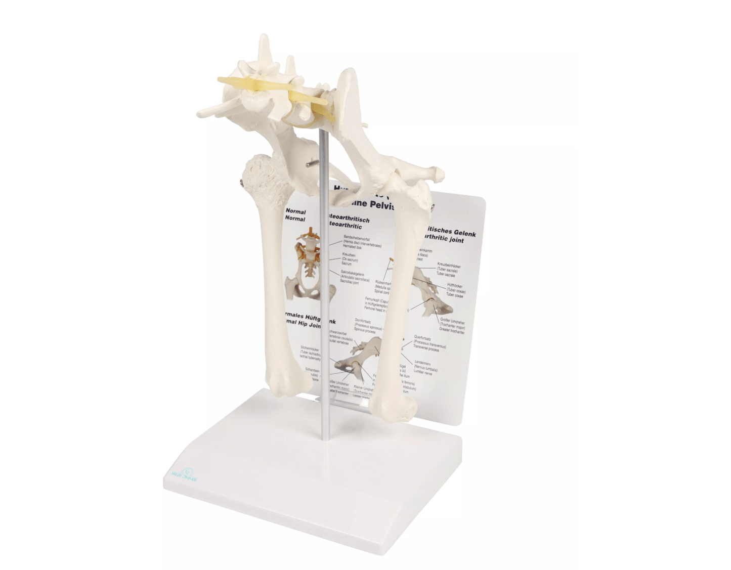
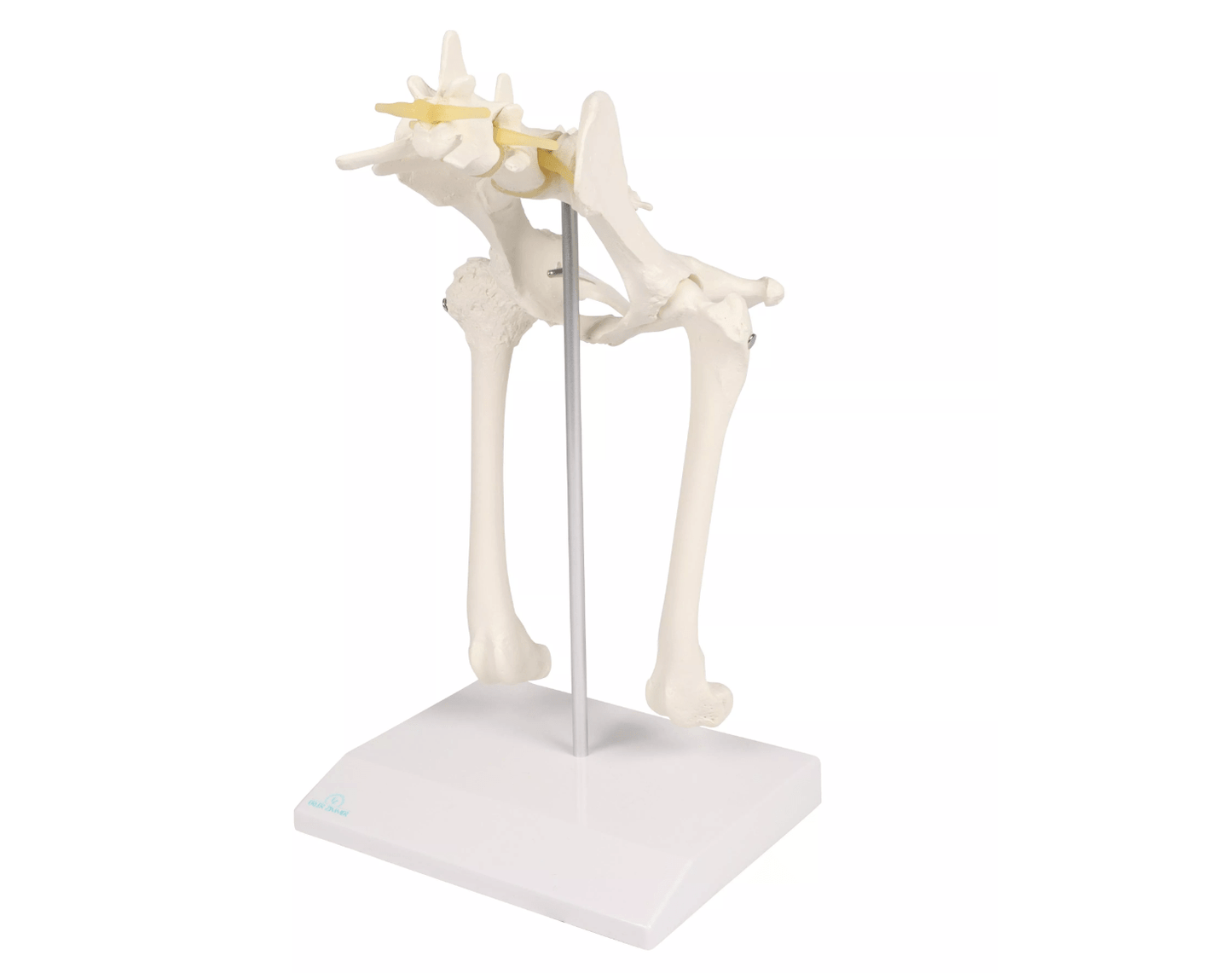
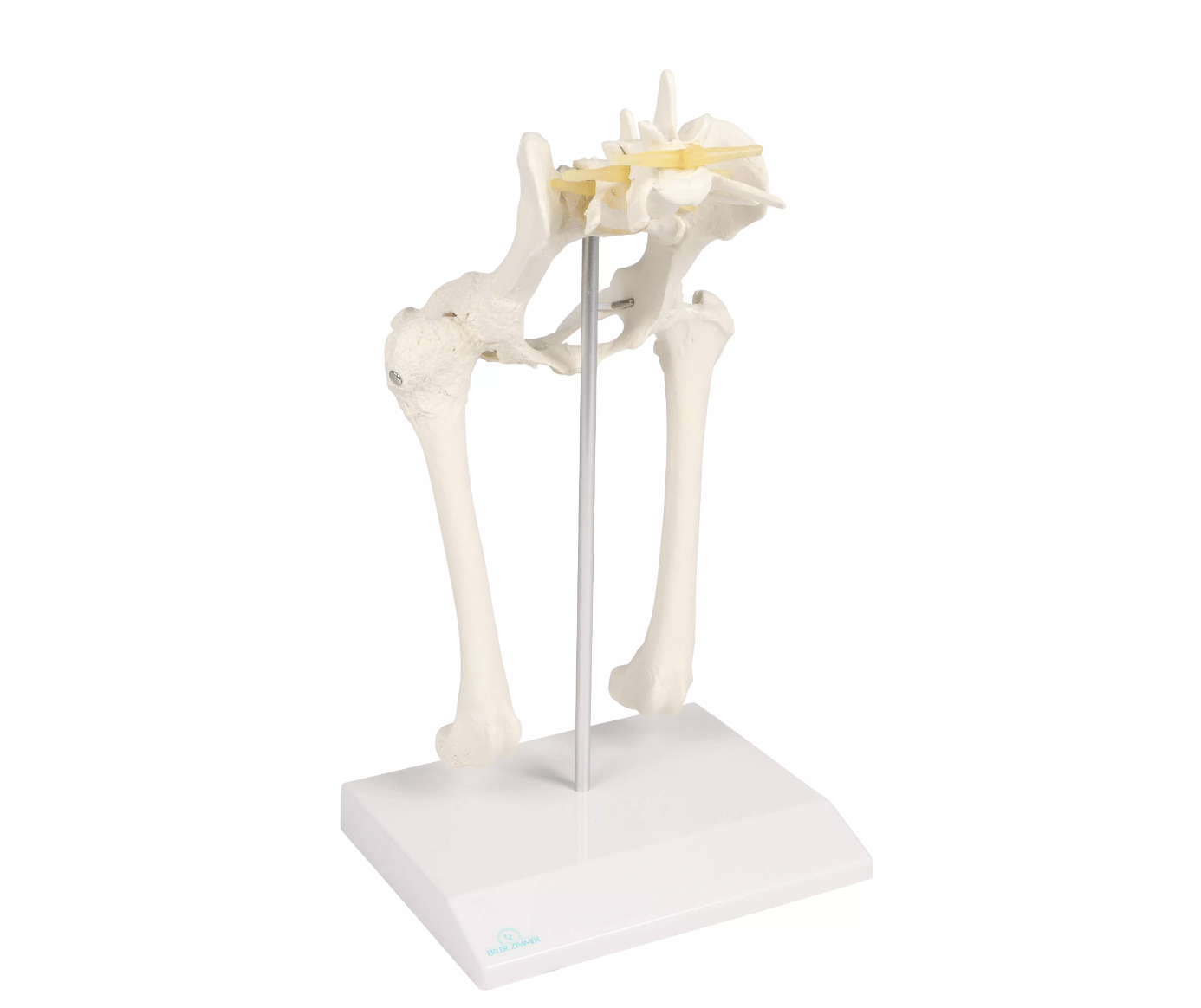
The medium-sized Canine Hip Joint Model provides a detailed and realistic representation of a dog's hip anatomy, including both healthy and arthritic bones. The model allows for a thorough examination of the anatomical structures that make up the hip joint and provides a vivid representation of the changes that can occur in arthritic diseases. Ideal for veterinary training and the analysis of hip problems in dogs, such as dysplasia or osteoarthritis.
Features and functions:
-
Life-size representation: The model shows the hip joint of a dog in natural size to enable a precise examination of the hip structure and its changes.
-
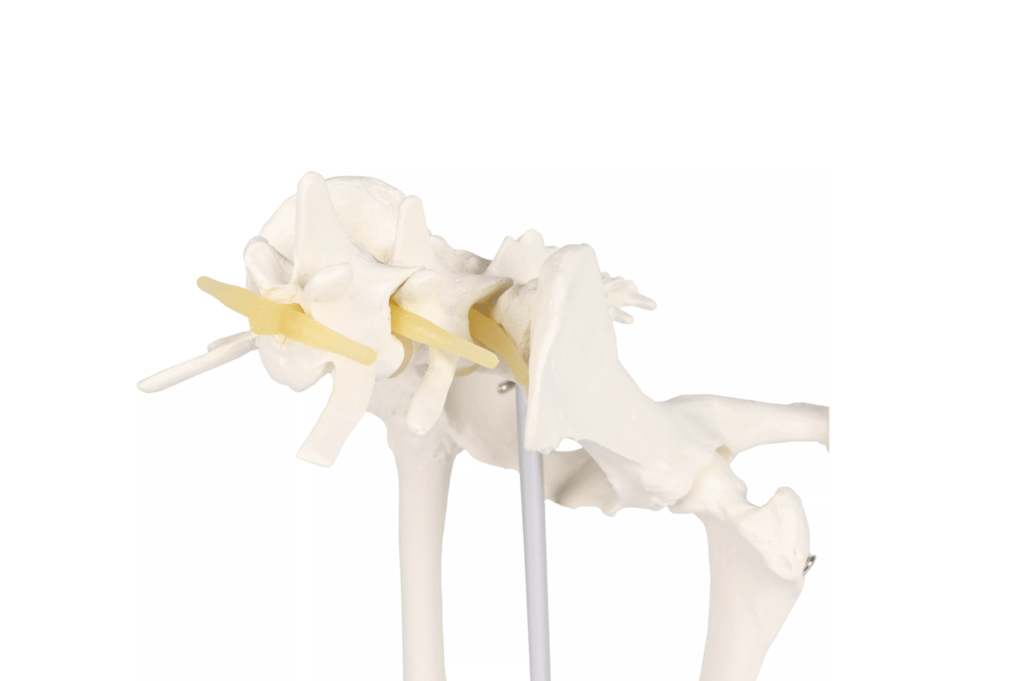
Healthy and arthritic bone: The model contains both healthy bone and arthritic bone, providing a vivid representation of the degeneration and effects of hip osteoarthritis.
-
Important anatomical structures: The model shows the important structures of the hip joint, including the corpus ilii , greater trochanter , femoral head in the acetabulum , disc herniation , femoral neck , nerve , sacrum and tertius trochanter .
-
Movable thigh: The thigh is movable and can be removed, allowing a detailed examination of the joint movements and an analysis of the functioning of the hip joint.
-
Precise depiction of degeneration: The depiction of healthy and arthritic bone enables a detailed examination of the symptoms and treatment options for hip osteoarthritis and hip dysplasia.
Advantages:
-
Realistic representations: The model provides a detailed and precise visualization of the canine hip joint, making it an excellent learning tool for veterinary students and veterinarians.
-
Differentiation between healthy and arthritic bones: By visualizing healthy and arthritic bones, students and professionals can better understand and analyze the effects of joint diseases.
-
Promotes understanding of hip problems in dogs: The model supports the understanding of hip dysplasia and osteoarthritis as well as their diagnosis and treatment options in veterinary medicine.
-
Movable and removable parts: The removable thigh allows for a more detailed examination of joint movements and the effects of arthritic changes.
Use and application:
The Canine Hip Joint Model is ideal for use in veterinary medicine, especially in training centers, universities, and clinics dedicated to the diagnosis and treatment of hip problems in dogs. It is excellent for analyzing hip dysplasia, osteoarthritis, and other joint diseases, as well as for teaching about hip anatomy in animals.
Delivery information:
PZN: " "
WEEE number:
Assistive device number:
Manufacturer:
Erler-Zimmer GmbH & Co. KG
Hauptstrasse 27
77886 Lauf
Deutschland
Tel: +49 07841 / 67191-0
Email: info@erler-zimmer.de
VAT ID number: Achtung! Medizinisches Ausbildungsmaterial, kein Spielzeug. Nicht geeignet für Personen unter 14 Jahren. Attention! Medical training material, not a toy. Not suitable for persons under 14 years of age.