SKU: SKU: EZV109
Erler-Zimmer Disc head model
Verpackungseinheit :
Couldn't load pickup availability


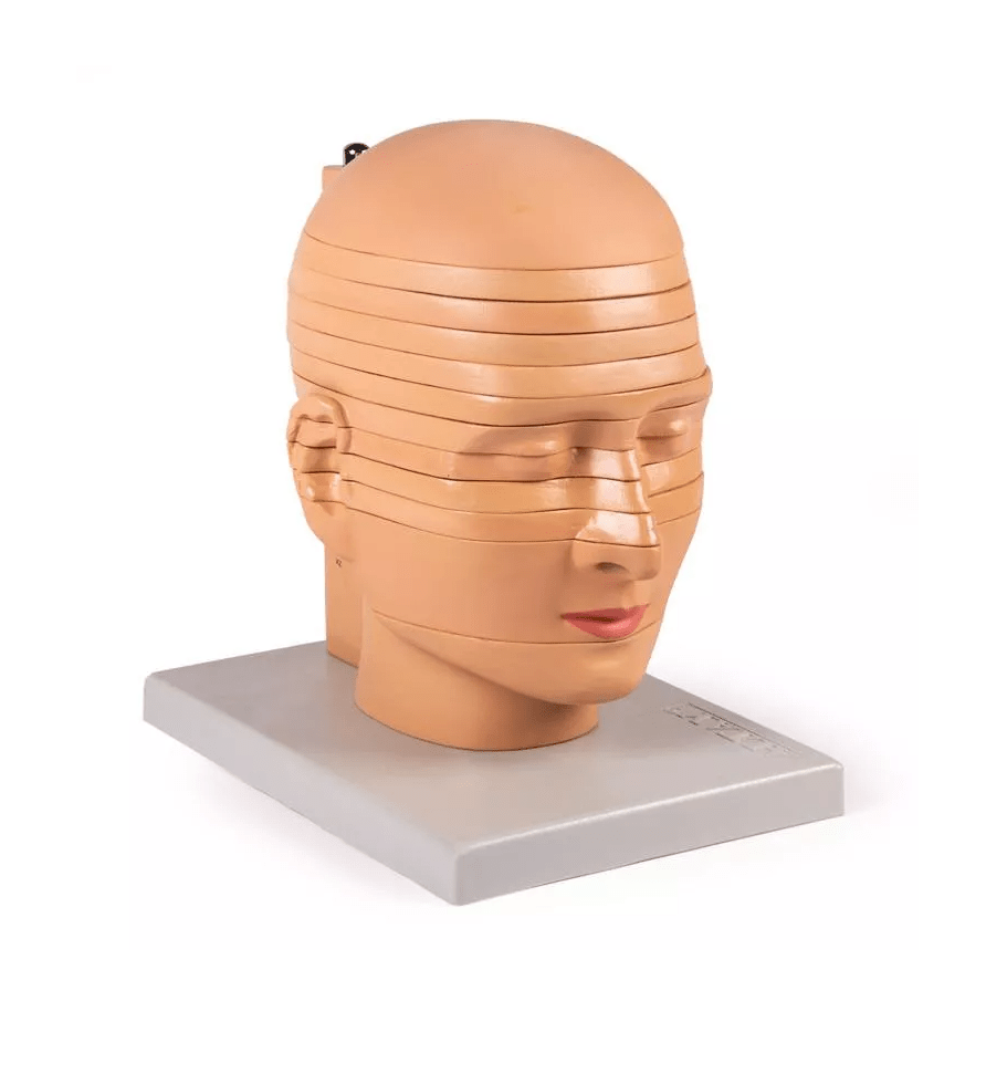
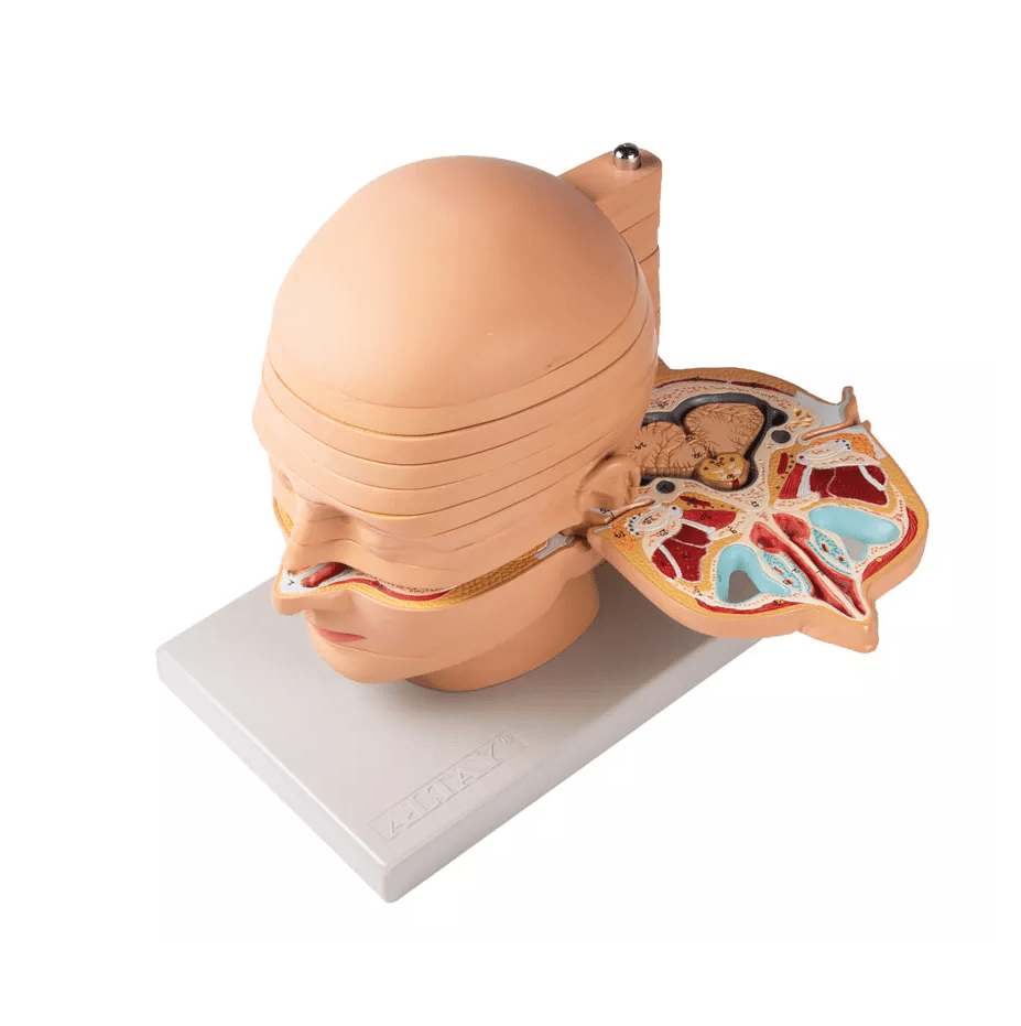
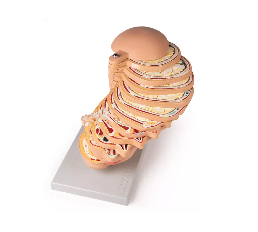
The Slice Head Model is a life-size anatomical model specifically designed to illustrate the workings of computed tomography (CT) and magnetic resonance imaging (MRI). This model is cut into 12 horizontal slices, allowing for detailed viewing of the various anatomical structures of the head. Each individual slice can be rotated or removed for precise examination of the intricate anatomical details. Ideal for medical students, trainees in radiology and anatomy, and anyone seeking an in-depth understanding of human anatomy.
Features and functions:
-
Life-size representation: The model shows the human head in natural size, so that all relevant structures are shown in detail and in the correct proportions to one another.
-
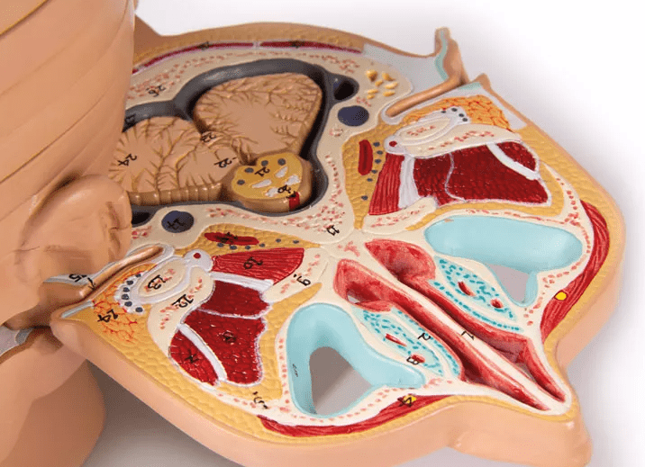
Horizontal slices: The head is cut into 12 horizontal slices, which allow each layer to be viewed individually and thus better understand the detailed structure of the brain and adjacent structures.
-
Rotatable and removable discs: Each disc can be rotated and removed to allow for even more detailed examination and better visualization of the layered structures of the head.
-
Anatomical details: All relevant structures, including muscles, cerebral gyri and sulci, are carefully numbered and described in detail in the included KeyCard.
-
Applications: Ideal for demonstrating the functionality of CT and MRI imaging as well as for studying the anatomical structures of the human head.
Advantages:
-
Practical learning aid: The model offers an excellent opportunity to learn and understand the structures of the head and the layering of tissues through imaging techniques such as CT and MRI.
-
Detailed anatomy: It precisely illustrates the different layers of the head, allowing a deeper insight into the anatomy, from the muscles to the brain.
-
Ideal for teaching and training: Perfect for medical schools, universities and training centers to promote understanding of brain structures, imaging techniques and their application.
Use and application:
The disc head model is ideal for medical students, radiologists, neurologists, and anyone interested in the anatomy of the human head and the workings of modern imaging techniques. It can be used in teaching units, practical exercises, and as a visual aid for research and diagnosis.
Delivery information:
PZN: " "
WEEE number:
Assistive device number:
Manufacturer:
Erler-Zimmer GmbH & Co. KG
Hauptstrasse 27
77886 Lauf
Deutschland
Tel: +49 07841 / 67191-0
Email: info@erler-zimmer.de
VAT ID number: Achtung! Medizinisches Ausbildungsmaterial, kein Spielzeug. Nicht geeignet für Personen unter 14 Jahren. Attention! Medical training material, not a toy. Not suitable for persons under 14 years of age.